
Page 27 - PlasticosVol4
P. 27
V O L 4 2019 I S S U E
flap by dissecting the greater omentum from Tip Case 2
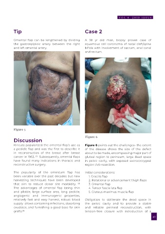
Omental flap can be lengthened by dividing A 38 yr old man, biopsy proven case of
the gastroepiploic artery between the right squamous cell carcinoma of natal cleft/spina
and left omental artery. bifida with involvement of sacrum, anal canal
and rectum.
Figure 2 Figure 3
Figure 2 shows the defect in the peritoneum flap by dissecting the greater omentum from
which was extended and omentum was the great curvature of the stomach, pedicled
exposed, separated, following which the entire by the right gastroepiploic artery, the perfusion
omentum was raised based on the right is secured by the arc of Barkow.
gastroepiploic vessels, perfusion secured by
the arc of Barkow. Figure 5
Figure 3 The omental flap was brought out of Figure 6
the peritoneal defect and an intraperitoneal Discussion
underlay technique utilizing a bilayer prosthetic Kiricuta popularized the omental flap’s use as Figure 6 points out the challenges- the extent
mesh was utilized, over which the peritoneum a pedicle flap and was the first to describe it of the disease shows the size of the defect
was closed. in reconstruction of the breast after breast about to be made, encompassing major part of
cancer in 1963. Subsequently, omental flaps gluteal region to perineum, large dead space
[1]
Figure 4 Omentum was spread over whole have found many indications in thoracic and in pelvic cavity, with exposed sacrococcygeal
of the defect which was covered with split reconstructive surgery. region i/v/o resection.
thickness graft.
The popularity of the omentum flap has Initial considerations:
Figure 5 Follow up picture. been variable over the past decades but new 1. Gracilis flap
harvesting techniques have been developed 2. Rotational or advancement thigh flaps
Technique that aim to reduce donor site morbidity. [2] 3. Omental flap
The omentum was identified and the The advantages of omental flap being thin 4. Tensor fascia lata flap
mobilization was begun by dissecting the and pliable, large surface area, long pedicle, 5. Gluteus maximus muscle flap
omentum along the avascular embryologic angiogenic and immunogenic properties,
fusion plane along the transverse colon. The relatively fast and easy harvest, robust blood Obligation to obliterate the dead space in
lateral splenic attachments and short gastric supply allows containing infections, absorbing the pelvic cavity and to provide a stable
vessels were divided to pedicle the omental flap exudates, and furnishing a good base for skin and reliable perineal reconstruction, with
on right gastroepiploic vessels. Harvesting the Figure 4 grafts. [3] tension-free closure with introduction of a
26 27